May 16, 2026/
No Comments
Gum disease starts quietly. There is no sharp pain, no dramatic swelling, nothing that screams emergency. For most people, the...


Gum disease starts quietly. There is no sharp pain, no dramatic swelling, nothing that screams emergency. For most people, the...
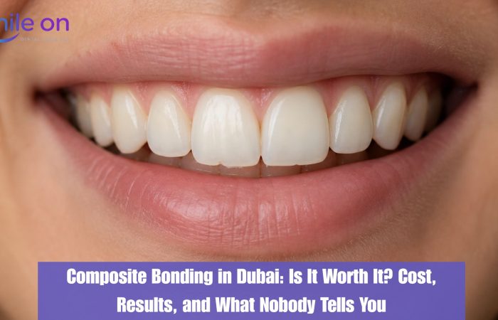
Most patients who walk into a cosmetic dental consultation in Dubai are already thinking about veneers. They have seen the...
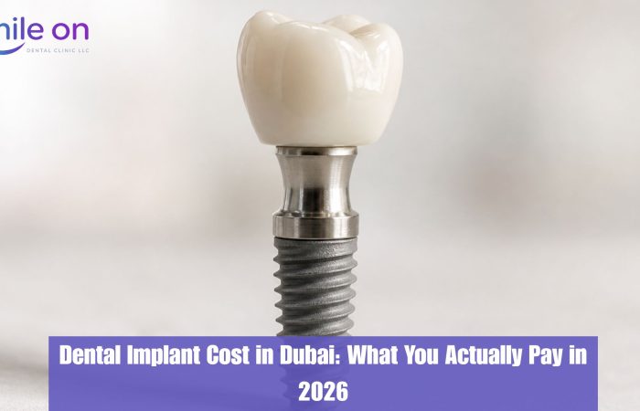
If you have been searching for dental implant prices in Dubai, you have probably seen numbers ranging from AED 1,500...